片側顔面痙攣の治療は、ボトックス局所注射、根治手術の2通りがあります。
通常は細い動脈が顔面神経に接触することが原因ですが、まれに太い血管、脳腫瘍が接触している場合があります。
正確な治療を行うために3D診断を事前に行います。
Treatment choice consists of two methods. For patients against surgery, Botox injection will be the first choice. However, the effect diminishes within 3 months and requires repeat injections. On the other hand, microvascular decompression can lead to complete cure. 3D imaging of the nerve and vessels is very helpful to understand the specific patient conditions which contributes to safer and effective surgery.
私たちの脳神経外科チームは、片側顔面けいれん(HFS)および三叉神経痛(TGN)に対する微小血管減圧術(MVD)に豊富な経験を有し、国内外から多くの患者さんを受け入れています。年間150〜200例のMVDを行っており、海外から日本へ手術を受けに来られる方も年々増加しています。世界水準の医療を提供すると同時に、国際患者を安心して受け入れられる体制を整えていることが大きな特徴です。
井上卓郎・後藤幸大・山岸正之
Our neurosurgical team has extensive experience in microvascular decompression (MVD) for both hemifacial spasm (HFS) and trigeminal neuralgia (TGN), performing approximately 150–200 procedures each year. We regularly welcome patients from around the world who choose to travel to Japan for surgery. With a well-established system for international patient care, we provide world-class treatment in a safe and supportive environment. Moreover, compared to many other countries, the cost of MVD in Japan is substantially lower—especially with the current exchange rate—making our care both highly specialized and cost-effective.
Takuro Inoue, M.D., Ph.D.; Yukihiro Goto, M.D., Ph.D.; Masayuki Yamagishi, M.D.
三叉神経痛で確立した3D診断を顔面痙攣にも応用し、鍵穴手術で手術を行います。典型的な場合は小切開、数時間での手術が可能です。従来から行われていたスポンジを神経と血管の間にはさむ方法ではなく、血管を完全に神経から離すことにより、治癒率を上げて、再発率を下げることが可能です。
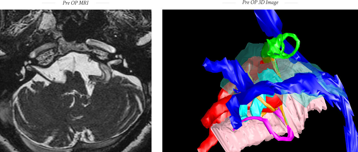
MR and CT imaging with 3D reconstruction are used to diagnose the cause of hemifacial spasm, which is usually an artery compressing the facial nerve, but may be due to other causes of compression.
Microvascular decompression for hemifacial spasm is performed through a key hole with small skin incision. Transposition techniques (sling or bridge technique) is used instead of the interposition technique to avoid failure and recurrence.

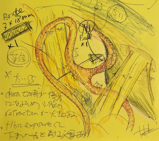
ブリッジテクニックを用いることにより確実な神経減圧が行えます。
Bridge technique can provide secure nerve decompression.
Bridge technique for hemifacial spasm with vertebral artery involvement.
Inoue T, Shitara S, Goto Y, Arham A, Prasetya M, Radcliffe L, Fukushima T.
This beautiful lady came from Hawaii, USA.
She suffered from hemifacial spasm on the right side for 9 years and was forced to quit her career as a school teacher due to HFS.
The 3D imaging revealed the anterior inferior cerebellar artery compressing the root entry zone of the facial nerve. This 3D imaging contributed to her successful MVD.
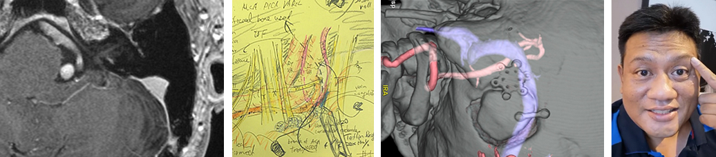
She came from Poland for the first MVD.
The common trunk of the anterior and inferior cerebellar artery (APC) was identified as a responsible vessel with 3D imaging. The offending artery was transposed using our Teflon Bridge Technique. She became spasm free immediately after surgery.
The APC and the Bridge Technique are described in our article.
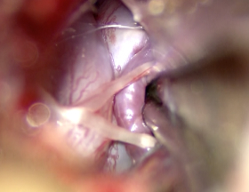
Operative findings
Pre OP
Post OP
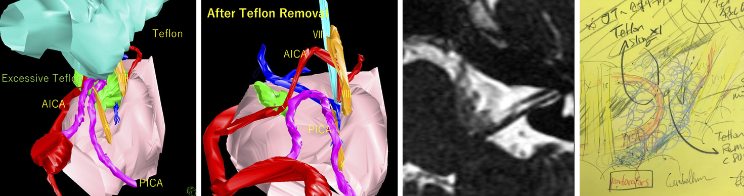
He is a physician in Nevada, USA, and suffered from hemifacial spasm on the left side for 5 years.
His first MVD was performed in Pittsburgh, Pennsylvania, which failed to improve the HFS. His HFS worsened after the first surgery.
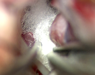
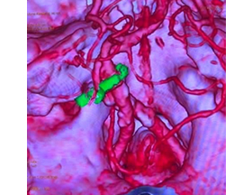
3D imaging created at Koto Memorial Hospital disclosed a massive Teflon (green object in 3D video) inserted between the facial nerve and the responsible artery. The surgery was performed to remove the excess Teflon (white material in OP video) to obtain sufficient decompression of the nerve. His HFS disappeared immediately after the surgery.

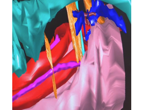
Pre - operative 3D imaging
The Teflon felt was removed
This patient had prior MVD with interposition technique.
HFS persisted due to too much Teflon inserted between the nerve and the compressing artery (microvascular recompression).
After removal of the excess of Teflon felt and appropriate transposition of the artery with sling technique, her symptoms were cured immediately after surgery.
Too much Teflon is compressing the facial nerve.
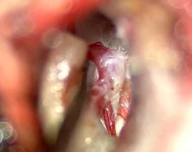
Teflon was removed and the compressing artery is exposed.
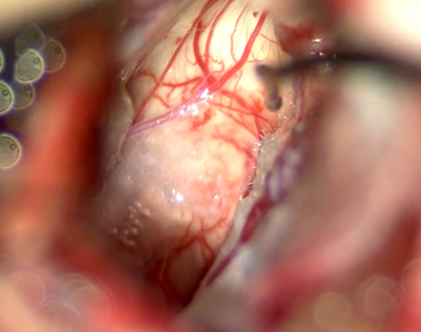
The artery is now transposed with the sling technique. The REZ of facial nerve is free from any kind of compression.
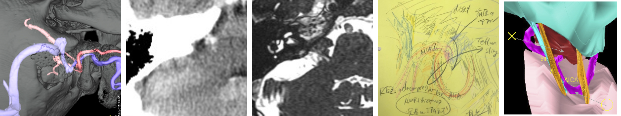
He underwent his 1st MVD in a local hospital in Singapore. The site of craniotomy made the procedure difficult, resulting in no improvement of HFS.
Our 3D imaging study revealed 4 arteries, both side of the vertebral arteries, the posterior inferior cerebellar artery, and the anterior inferior cerebellar artery, were involved in neurovascular compression. The vessels were securely lifted with a Teflon bridge away from the REZ.
3D imaging showed multiple vascular compression.
The arteries were elevated from the REZ.
The post OP CT showed that the vessels were securely elevated with a Teflon bridge (green).
He is so happy with spasm free after 2nd MVD.
67 F
Hemifacial spasm on the left side for the last 12 years.
Surgery at other hospitals failed to relieve her facial spasm.
We precisely evaluated using 3D imaging and performed microvascular decompression, resulting in successful relief of her symptom.
These large vessels require meticulous decompression technique.
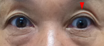
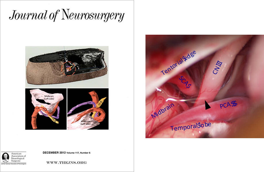
62 F
Paroxysmal diplopia when stressed or speaking with others, lasting ~10-20 sec. Neurological finding revealed paroxysmal downward deviation of the left eye when the spell occurs. 3D imaging showed the left oculomotor nerve pinched by two different arteries. Microvascular decompression transposed these arteries, leading to resolution of her symptom.
※要予約※Reservation required.
| 湖東記念病院 Koto Memorial HospitalWebSite |
平日 Weekdays |
午後 p.m. |
| 学研都市病院 Gakkentoshi HospitalWebSite |
第3土曜日 Third Saturday |
午前 a.m. |
| 蘇生会総合病院 Soseikai HospitalWebSite |
第3土曜日 Third Saturday |
午後 p.m. |
| 湖東記念病院 Koto Memorial HospitalWebSite |
日本、滋賀 Shiga, Japan |
| 蘇生会総合病院 Soseikai General HospitalWebSite |
日本、京都 Kyoto, Japan |
| 志太記念脳神経外科 Shida Memorial Neurosurgery ClinicWebSite |
日本、静岡 Shizuoka, Japan |
| Andalusia HospitalWebSite | Alexandria, Egypt |
| National Brain Center HospitalWebSite | Jakarta, Indonesia |
湖東記念病院脳神経外科主任部長
日本脳神経外科学会専門医